Vascular Disorders
A variety of conditions can affect the blood vessels of the brain and spinal cord. Although many are found before they become symptomatic, others only come to attention after they have caused bleeding in the brain. When bleeding in the brain has occurred, decisive action is needed. Dr. Hatef has extensive experience with vascular conditions including aneurysms, arteriovenous malformations and fistulas, strokes, and moya-moya disease.
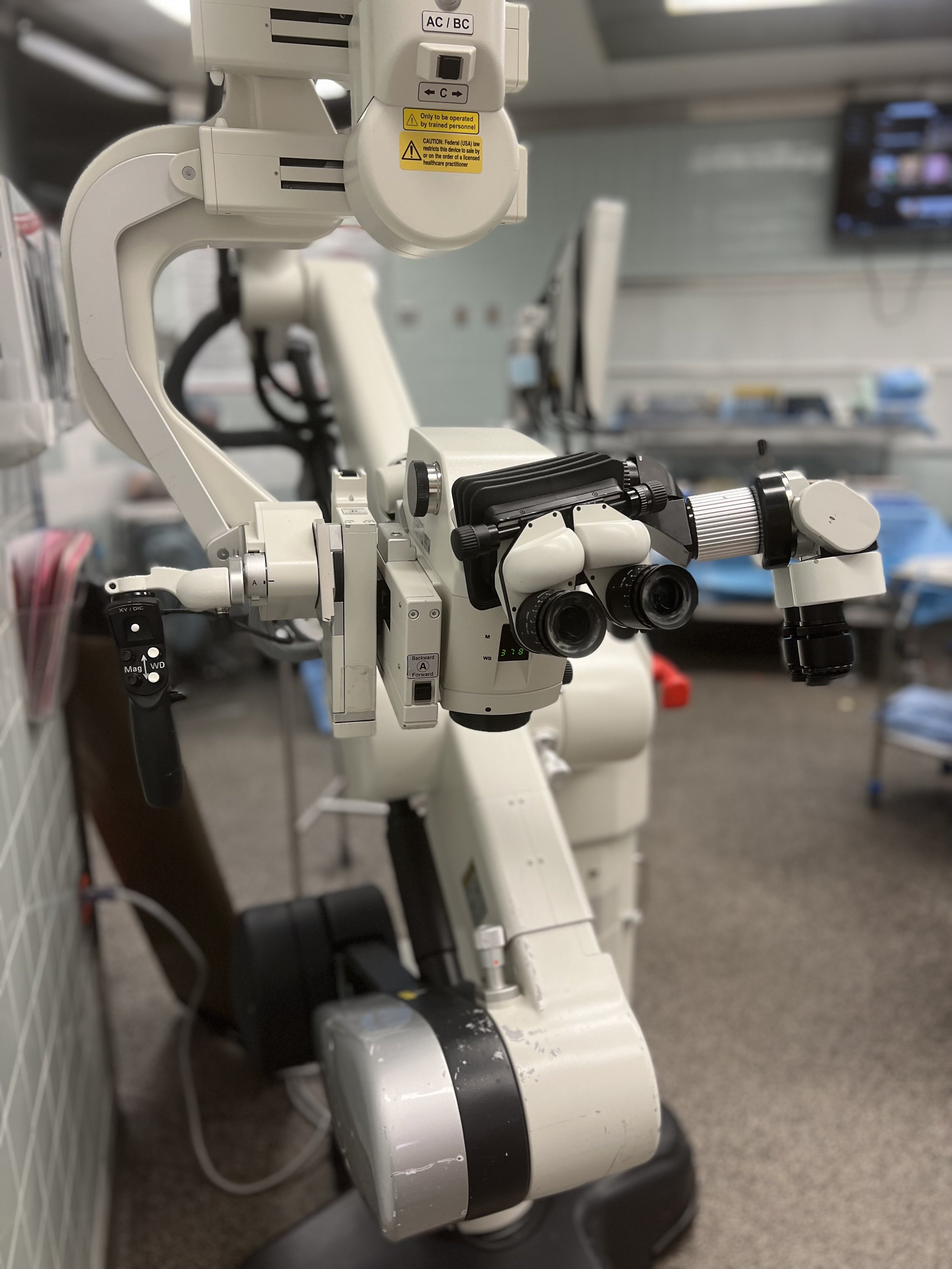
Dr. Hatef uses the operative microscope for all vascular procedures to ensure optimal visualization.
Most neurovascular conditions are treated by a craniotomy - removing a window of bone to allow Dr. Hatef to identify and correct the abnormality. With advances in neuroanesthesia, surgical navigation, and use of the intraoperative microscope, modern neurovascular surgery is safe and effective.
For those patients who have suffered a rupture of their vascular disorder with intracranial bleeding, time is of the utmost importance. Emergency surgery may be needed to relieve pressure on the brain and prevent further bleeding. Identifying these lesions before they bleed is the best way to prevent damage to the brain.
Dr. Hatef is committed to a shared decision-making model of care in neurosurgery. He believes that with his help and advice, patients should have autonomy to decide which course of treatment is right for them. Please find more information about different vascular conditions in neurosurgery below.
Aneurysms
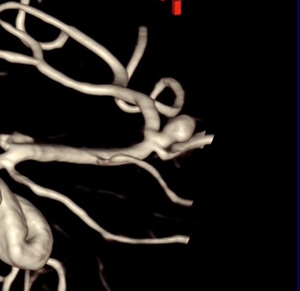
This 3-D angiogram shows a large aneurysm of the middle cerebral artery. This was treated with clip ligation.
Aneurysms are abnormal pouches in the walls of cerebral blood vessels. The arise from weakness in the layers of the vessel. Although they are typically small - usually less than 1 centimeter - when they rupture they can cause significant damage or even death. In his practice, Dr. Hatef treats two types of aneurysms: unruptured and ruptured aneurysms. For more information about treatments, click here.
Unruptured aneurysms
Unruptured aneurysms are usually diagnosed when a patient received CT or MRI scans for headache or trauma. These aneurysms are usually asymptomatic until they rupture. The goal of treatment is to safely occlude the aneurysm and prevent its rupture. Some aneurysms are amenable to endovascular techniques - a radiologist will guide a catheter into the aneurysm and use specialized devices to occlude it. Others require surgery for clip ligation. During an aneurysm clipping, Dr. Hatef will gently identify the aneurysm and use metal clips to ensure it can no longer fill with blood, preventing its rupture. Whether an aneurysm should be treated, and what kind of treatment it requires, depends on the specific characteristics of an individual aneurysm. For those with unruptured aneurysms, Dr. Hatef will take time to explain how risky the aneurysm appears and what he thinks is the best treatment course. Together, a plan will be developed to best deal with your aneurysm.
Ruptured aneurysms
A ruptured aneurysm is a true neurosurgical emergency. Many patients will not survive the initial rupture, and many of those that do will suffer permanent brain damage. The best way to prevent a rupture is to identify and treat aneurysms beforehand. For patients that suffer a ruptured aneurysm, the aneurysm will need to be urgently treated with clipping or endovascular techniques. It is common for these patients to spend significant time in intensive care before rehabilitation. Dr. Hatef follows with his patients at every step to help them through this frightening situation.
Arteriovenous malformations
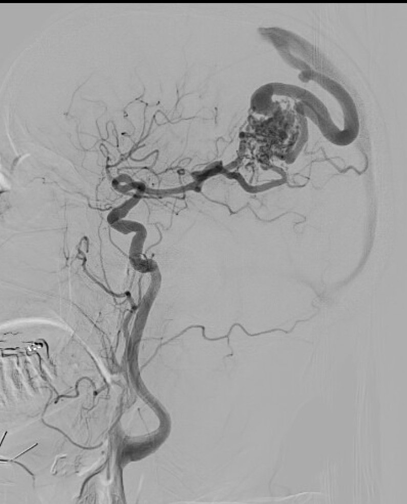
This large AVM is fed through abnormal vessels from the middle cerebral artery.
Arteriovenous malformations (AVM) are abnormalities of the blood vessels on the surface of the brain. In a normal brain, blood flows gently from arteries to capillaries and veins. In an AVM, blood rushes from arteries into a tangle of vessels called the nidus. Because the pressure is so high and the blood flow so abnormal, AVM are at risk of rupture, causing bleeding into the brain.
Similar to aneurysms, AVM can be diagnosed before or after rupture. Most AVM are at a significant risk of rupture and should be treated after diagnosis.
Treatments for AVM include surgery, radiation treatments, and endovascular treatments. If you have been diagnosed with an AVM, Dr. Hatef and the team of radiologists and radiation oncologists will work to provide the best treatment for your specific malformation.
Stroke
Stroke is a common disease and significant cause of disability in the United States. Strokes can either be ischemic, meaning blood flow to brain has stopped, or hemorrhagic, meaning bleeding has occurred within the brain.

A hemorrhagic stroke in the left cerebellum.
Hemorrhagic strokes can be due to underlying conditions such as aneurysms or malformation, but they can also arise spontaneously. The most important issue in treating a brain hemorrhage is to relieve the pressure inside the head, or intracranial pressure.
Although intracranial pressure can be treated with medication, surgery is often needed to remove the blood clot and relieve the pressure on the normal brain. If you have been diagnosed with a hemorrhagic stroke, Dr. Hatef will determine if surgery is required and work with a team of neurologists to treat your condition.
Ischemic strokes occur when blood flow to the brain is cut off. Symptoms typically develop within seconds. If strokes are diagnosed quickly enough, treatments with medical therapy and surgery can greatly improve neurologic outcome.

A large ischemic stroke in the left hemisphere.
In patients with large strokes, swelling within the stroke can compress the surrounding normal brain. Surgery is often needed to decompress the stroke and prevent further brain damage. A procedure called a hemicraniectomy involves removing a large window of the skull bone to allow the pressure inside the head to decrease. Patients with strokes this large require admission to intensive care and close coordination among the surgical, neurology, and intensive care team.
Patients with smaller strokes and carotid artery stenosis are often good candidates for carotid endarterectomy to prevent future strokes. To learn more about procedures Dr. Hatef offers to prevent stroke, please click here.
Other Conditions
Moya-Moya disease results when the blood vessels of the brain slowly start to constrict, leading to decreased blood to the brain. Dr. Hatef offers a surgical procedure to reverse the loss of blood vessels and help prevent strokes and hemorrhages.
Arteriovenous fistula are abnormal connections between brain arteries and veins. Most can be treated by our radiology colleagues with catheter-based techniques. For those that are not treatable with endovascular means, Dr. Hatef offers surgical disconnection of the fistula.
Cavernous malformations are abnormal collections of small blood vessels. They can cause problems with bleeding or seizures. Dr. Hatef performs craniotomies to remove cavernous malformations to prevent bleeding and help with seizure control.
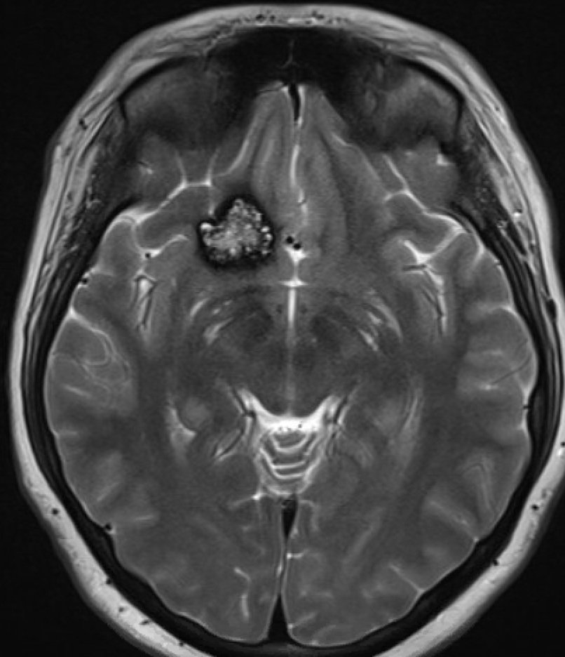
A cavernous malformation in the right frontal lobe. This was removed with a small, well-hidden incision in the eyebrow.