Chiari Malformations and CSF Disorders
Cerebrospinal fluid (CSF) is produced continually by the brain and bathes the brain and spinal cord. It surrounds the nerves and cushions them. Dr. Hatef treats a variety of disorders involving the CSF, including chiari malformations, syringomyelia, hydrocephalus, pseudotumor cerebri, and normal pressure hydrocephalus.
Chiari malformations and syringomyelia

A chiari malformation. The cerebellum is displaced into the spine, compressing the spinal cord.
Chiari malformations are abnormalities of the cerebellum, the portion of the brain near the base of the skull. Originally named for Dr. Hans Chiari over 100 years ago, our understanding of this condition has expanded greatly in the past few decades.
The cerebellum is normally entirely within the skull cavity. However, in Chiari malformation, the cerebellum is displaced into the spinal canal. This can result in compression of the brainstem or spinal cord, or resistance to the normal flow of CSF.
A Chiari malformation is typically diagnosed due to headaches. The headaches are often severe, pounding, and over the back of the skull and upper neck. In many patients the headaches are worsened by bearing down, such as during lifting or bowel movements. For patients with severe headaches, a procedure called a Chiari decompression can provide excellent relief. The lower-most part of the skull is carefully removed, relieving the compression of the brain and allowing the cerebellum to return to a more normal position.

Syringomyelia. There is a large fluid-filled cyst within the spinal cord.
A related condition is syringomyelia. A syrinx is an abnormal collection of fluid within the spinal cord. These are thought to develop due to resistance to the flow of CSF and are commonly seen in patients with Chiari malformations. Syrinxes can cause numbness, tingling, or weakness and are a sign that a Chiari malformation is severe. Although the syrinx is down in the spinal cord, in patients with Chiari malformation, the problem is still at the base of the skull. Chiari decompression will improve the syrinx and prevent damage to the spinal cord.
Hydrocephalus
Hydrocephalus is the medical term for fluid on the brain, or fluid within the ventricles. The ventricles are fluid-filled spaces in the center of the brain where CSF is normally produced. In most people the CSF is produced and flows throughout the brain. However, in individuals with hydrocephalus, the natural flow of CSF is restricted. This causes a backup of fluid and an increase in pressure.
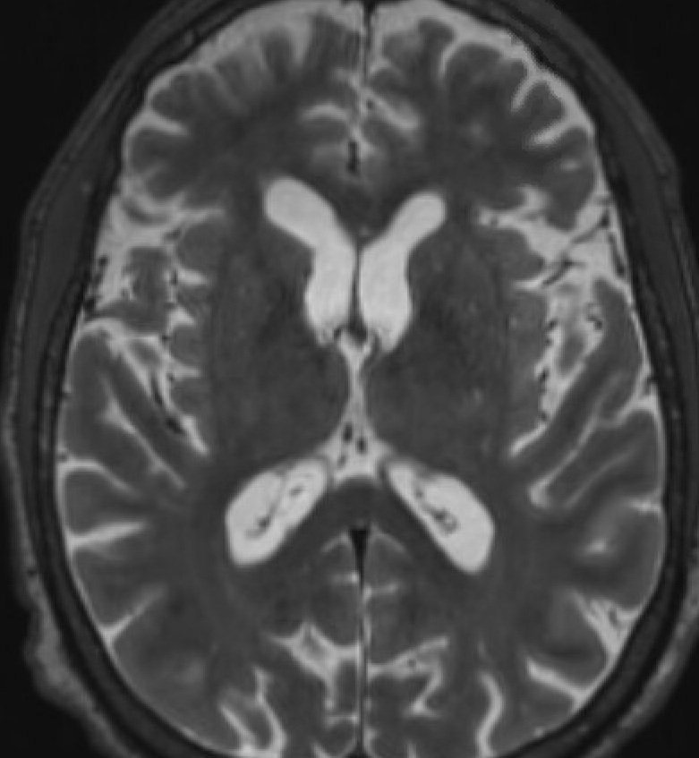
Normal ventricles are the white cavities within the brain.

Severe hydrocephalus with dramatic enlargement of the ventricles.
The treatment of hydrocephalus is usually a shunt procedure. A small tube is placed into the ventricle to drain the fluid. Most common it is drained down into the abdomen where it is absorbed without issue. Most shunt procedures are short surgeries with just one day in the hospital. Dr. Hatef believes in being as minimally invasive as possible, with small incisions, minimal clipping of hair, and absorbable sutures.
Pseudotumor cerebri
Pseudotumor cerebri, also called idiopathic intracranial hypertension (IIH), is a condition that is being more commonly diagnosed. Patients with pseudotumor have elevated pressure in the head, but unlike patients with hydrocephalus, they do not have enlargement of the ventricles. Often patients have severe headaches and can have vision loss or even blindness. Although pseudotumor cerebri is usually managed with medication, for those who continue to have symptoms, Dr. Hatef offers shunting procedures to help relieve pressure.
Normal Pressure Hydrocephalus (NPH)
Normal pressure hydrocephalus (NPH) is a common condition as patients age. Common symptoms include difficulty walking, cognitive changes, and urinary incontinence. Dr. Hatef works closely with a team of neurologists to diagnose this condition - typically with MRI scans and a lumbar puncture or spinal tap. Once NPH is diagnosed, insertion of a CSF shunt often relieves symptoms.