Brain Tumors
Brain tumors are growths that arise within the brain or its surrounding coverings. They may be benign or malignant depending on the specific diagnosis. A thorough neurologic exam and detailed magnetic resonance imaging (MRI) scan are critical to accurately diagnosing a brain tumor.
Once a brain tumor has been diagnosed, surgery is often required. Tumors are removed via a craniotomy: an incision is made in the scalp and a window is made in the skull to access the tumor. Tumors are then carefully removed from the surrounding normal brain. Dr. Hatef does his best to make this operation as least invasive as possible - minimal hair is removed, and the scalp is closed with absorbable sutures instead of traditional staples.
How a brain tumor will affect your life greatly depends on the specific diagnosis. Information is available below regarding the most common brain tumors. Please click here to learn more about a craniotomy procedure for your brain tumor.
Meningioma

A meningioma is seen over the right orbit, compressing the brain and distorting the bone of the eye socket.
Meningiomas are benign tumors that arise from the covering of the brain - the meninges - instead of the brain itself. The are often slow growing tumors. Meningioma can arise anywhere in the brain, and the symptoms they cause depend on what structures are nearby. They can cause headaches, vision loss, weakness, numbness, and seizures.
The goal of surgery is the remove the entire meningioma without injuring the normal brain. With many meningiomas, including the tumor pictured here, skull base surgery techniques are required to avoid any injury to the brain. Dr. Hatef is experienced in the safe approach and resection of skull base meningiomas and employs the most advanced techniques to protect his patients.
Glioblastoma and glioma

A glioblastoma is present in the right temporal lobe. This patient presented with seizures and underwent a full resection.
Glioblastomas and gliomas arise from cells within the brain. The range in aggressiveness, and glioblastoma is commonly referred to as brain cancer. Although glioblastomas are typically not curable with surgery, removing the maximum amount of tumor safely will prolong survival and increase quality of life.
Dr. believes in a compassionate and personal approach to patients with glioblastoma. He works closely with oncologists to develop a plan specific to an individual patient.
Vestibular Schwannomas

A vestibular schwannoma is seen on the right side of the brain, compressing and distorting the brainstem.
Vestibular schwannoma, also known as acoustic neuroma, are benign tumors that arise from the nerve responsible for hearing and balance. They often come to attention based of symptoms of hearing loss, ringing in the ear, and balance trouble. Although they are benign, they can grow to large size and compress the normal brain, leading to sometimes dramatic symptoms.
Vestibular schwannomas are treated with surgery and/or radiation, depending on the size of the tumor. Through a small incision behind the ear, most tumors can be safely removed. As they are slow growing tumors. observation or radiation treatments are often reasonable, especially in older patients.
Metastases
The most common brain tumors are tumors that arise elsewhere in the body. The then spread, or metastasize, to the brain. The most common metastatic tumors are breast cancer, lung cancer, kidney cancer, and melanoma. When a patient has metastatic cancer, we typically know the type, which makes retrieving tissue for examination under the microscope less necessary. Treatment for metastatic brain tumors requires the input of neurosurgeons, radiation oncologists, and medical oncologists. Surgery is often needed to remove large tumors and relieve compression of the brain. Dr. Hatef works closely with a team of oncologists to determine which surgical, medical, and radiation treatments are right for your specific tumor.

There is a large amount of edema in the left parietal lobe associated with a metastatic lung tumor.
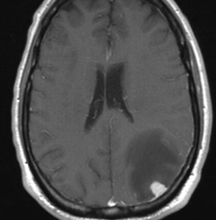
Although the tumor was quite small, it caused a significant amount of swelling in the surrounding normal brain.